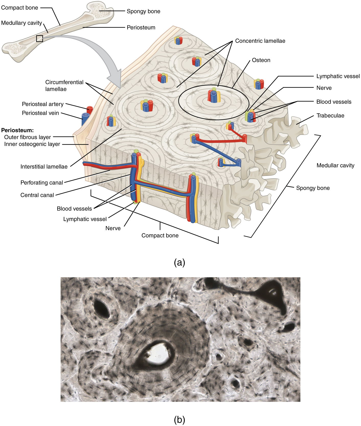
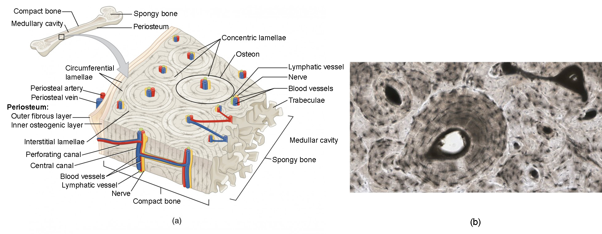
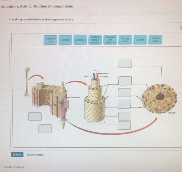
Anatomy and Histological Organization of Bone Label the structural features of compact bone. The osteocytes are arranged in concentric rings of bone matrix called lamellae little plates and their processes run in interconnecting canaliculi.
Bone Structure Anatomy And Physiology
Reduction refers to _____________.

. The inner osteogenic layer consists primarily of ________. The loss of bone tissue as you age. Start studying Art-labeling Activity.
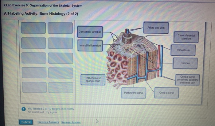
Examining the Histology of a Lymph Node a Tonsil and the Spleen. The differences between compact and spongy bone are best explored via their histology. Correct Help Reset Help Reset Interstitial lamellae Osteons Perforating canal Trabeculae of spongy bone Perforating fibers Periosteum Concentric lamellae Central canal Circumferential lamellae.
Compact bone is organized as parallel columns known as Haversian systems which run lengthwise down the axis of long bones. The structural unit of compact bone is the osteon an elongated cylinder oriented parallel to the long axis of the bone. Label the types of bone cells.
The bone would be stronger. Compact bone is dense so that it can withstand compressive forces while spongy cancellous bone has open spaces and. Exploring the Microscopic Anatomy of Compact Bone.
The diaphysis is the tubular shaft that runs between the proximal and distal ends of the bone. Functionally it assumes a significant mechanical role by the skeleton and represents a stock of mineral salts to mobilize for maintenance of calcium and phosphorus homeostasis. Most bones contain compact and spongy osseous tissue but their distribution and concentration vary based on the bones overall function.
A typical long bone showing gross anatomical features. BONES OF THE AXIAL AND APPENDICULAR SKELETON. A typical long bone shows the gross anatomical characteristics of bone.
Structure of a Long Bone. This photo shows a cross section through bone. Drag the labels onto the diagram to identify the structures found in compact bone.
Histology of Nervous Tissue Label the parts of a representative neuron. The Histology of Compact Bone Identify the microscopic structures of bone. Running down the center of each osteon is the central canal or Haversian canal which contains blood vessels nerves and lymphatic vessels.
Types of Bone Cells Learning Goal. Part A Drag the labels to identify the microscopic structures of bone. To learn the structures found in compact bone.
View Homework Help - API Lab Homework 7 from BSC 2085L at University of South Florida. The outer walls of the diaphysis cortex cortical bone are composed of dense and hard compact bone a form of osseous tissue. Art Labeling and Art-based Activity assignments are updated.
Classification of Bones by Shape. The structure of a long bone allows for the best visualization of all of the parts of a bone Figure 1. 10142016 API Lab Homework 7 28 Correct Artlabeling Activity.
Part A Drag the labels onto the diagram to identify the types of bone cells. Setting a broken bone back in its place. These columns are composed of lamellae concentric rings of bone surrounding a central channel or Haversian canal that contains the nerves blood vessels and lymphatic system of the bone.
Learn vocabulary terms and more with flashcards games and other study tools. Part A Drag the labels to the appropriate location in the figure. To learn the types of bone cells.
Anatomy and Histological Organization of Bone Label the structural features of compact bone. Bones are covered and lined by a protective tissue called periosteum. Start studying Art-labeling Activity.
10142016 APILabHomework7 APILabHomework7 Due1159pmonFridayOctober142016. Through the medullary spaces it. Study from the bone list or your textbook after you marked the drawings as instructed on page 6-2.
Most but not all features you are required to know are shown on the following pages. The Histology of Compact Bone. After you have studied the bones in lab label the drawings as a self-test.
The wider section at each end of the bone is called the epiphysis plural epiphyses which is filled internally with. Learn vocabulary terms and more with flashcards games and other study tools. Part A Drag the labels to the appropriate location in the figure.
Help Reset Apical portion of cell breaking down Golgi apparatus Secretory vesicles fusing with the plasmalemma Stem cell dividing to replace lost cells Holocrine secretion. Bone is the primary anatomical structure comprising of the human skeletal system. Loss of electrons in a chemical reaction.
Figure 631 Anatomy of a Long Bone. 41522 133 PM Week 1 Chapter 3 413 Correct Art-labeling Activity. The diaphysis and the epiphysis.
The central Haversian canal and horizontal canals perforating Volkmanns canals contain blood vessels and nerves from the periosteum. Learn vocabulary terms and more with flashcards games and other study tools. It protects several vital organs skull vertebrae and rib cage.
Structure of Compact Bone. Loss of bones through amputation. Each osteon is composed of concentric rings of calcified matrix called lamellae singular lamella.
Examining the Chemical Composition of Bone. Start studying Art-labeling Activity. The microscopic structural unit of compact bone is called an osteon or Haversian system.
Label the structures found in compact bone. Bone Markings Part 1. A long bone has two parts.
Mastering A P Chapter 6 Bones And Skeletal Tissues Flashcards Quizlet

2015 Pearson Education Inc Ppt Download
Solved Lab Exercise 9 Organization Of The Skeletal System Chegg Com
Bone Structure Anatomy And Physiology I

Art Labeling Activity Structure Of Compact Bone Diagram Quizlet
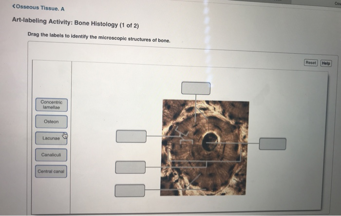
Solved Cou Osseous Tissue A Art Labeling Activity Bone Chegg Com

6 The Skeletal System Ppt Download
Solved Art Labeling Activity Structure Of Compact Bone Chegg Com
0 comments
Post a Comment